NC
Nasal cavity
Air-filled space in the nose for airflow and smell
RegionHead and Neck
SystemRespiratory System
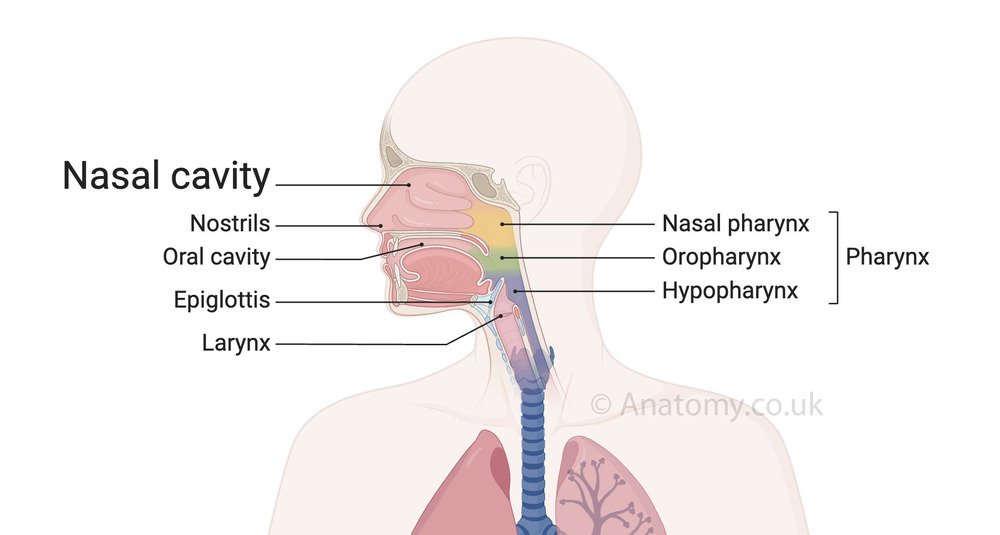
The nasal cavity is a hollow, air-filled space located within the nose and extending behind it. It is a crucial component of the respiratory system, playing an essential role in filtering, warming, and humidifying the air we breathe before it reaches the lungs.[8] The nasal cavity also houses the olfactory receptors, which are responsible for our sense of smell.
 This cavity is divided into two symmetrical halves by the nasal septum, a thin wall made of cartilage and bone. Each half of the nasal cavity opens to the nostrils at the front and connects to the throat at the back, forming a passage for airflow. The inner walls of the nasal cavity are lined with a specialized mucous membrane and tiny hair-like structures called cilia, which help trap dust, allergens, and other particles.
Due to its structure and functions, the nasal cavity plays a pivotal role not only in breathing but also in protecting the respiratory tract from harmful substances.[7] It works closely with the paranasal sinuses, the pharynx, and the larynx to maintain proper respiratory health. Additionally, its connection to the olfactory system highlights its importance in sensory perception.
Location
The nasal cavity is centrally located within the skull, just behind the external nose. It is positioned above the oral cavity, below the cranial cavity, and in front of the nasopharynx. Its boundaries include the frontal bone at the top, the hard palate at the bottom, and the ethmoid and sphenoid bones at the rear.[1] The nasal cavity opens externally through the nostrils and internally through the choanae, connecting to the nasopharynx.Anatomy
The nasal cavity is divided into two halves by the nasal septum, which consists of bone and cartilage. Each half features three bony projections called nasal conchae or turbinates (superior, middle, and inferior).[5] These structures create narrow passageways called meatuses, which help increase the surface area for air filtration and conditioning. The cavity is lined with a mucous membrane containing goblet cells that produce mucus, and cilia that trap and move particles out of the respiratory tract. The olfactory epithelium, located in the upper part of the cavity, is specialized for detecting odors. The nasal cavity also communicates with the paranasal sinuses, which are air-filled spaces within the skull, aiding in ventilation and drainage.[4]Structure of the Nasal Cavity
The nasal cavity is a complex structure divided into several distinct parts, each with a specific role in airflow and filtration.Nasal Septum
- Divides the nasal cavity into left and right halves.
- Made up of the vomer bone, perpendicular plate of the ethmoid bone, and cartilage.
Nasal Conchae (Turbinates)
- Three pairs of curved bony structures (superior, middle, and inferior) that project into the nasal cavity.
- Create narrow air passages called meatuses, increasing surface area and aiding in air conditioning.[6]
Nasal Meatuses
- Spaces formed beneath each concha.
- Serve as pathways for air and drainage from the paranasal sinuses and nasolacrimal duct.
Vestibule
- The anterior part of the nasal cavity, located just inside the nostrils.
- Lined with coarse hairs (vibrissae) that trap large particles.[3]
Olfactory Region
- Located at the roof of the nasal cavity.
- Contains the olfactory epithelium responsible for detecting smells.
Respiratory Region
- The largest part of the nasal cavity.
- Lined with ciliated pseudostratified columnar epithelium and goblet cells, which filter, humidify, and warm the incoming air.
Layers and Lining of the Nasal Cavity
The nasal cavity is lined with two primary types of tissue: 1. Respiratory Epithelium:- Pseudostratified columnar epithelium with goblet cells.
- Produces mucus that traps dust and pathogens while cilia transport them out.
- Located in the upper region for detecting odors.
- Contains sensory neurons and supporting cells.
Blood Supply and Nerve Innervation
Blood Supply:- Arterial supply from the sphenopalatine artery, anterior and posterior ethmoidal arteries, and facial artery.[2]
- Rich vascularization aids in warming inhaled air.
- Sensory nerves from the trigeminal nerve (V1 and V2 branches).
- Olfactory nerve (Cranial Nerve I) for smell detection.
Paranasal Sinuses and Their Connection
Frontal, Ethmoid, Sphenoid, and Maxillary Sinuses:- Connected to the nasal cavity via narrow openings.
- Aid in reducing skull weight, humidifying air, and enhancing voice resonance.
Openings and Connections
- Choanae: Connect the nasal cavity to the nasopharynx.
- Nasolacrimal Duct: Drains tears into the nasal cavity.
- Eustachian Tube Openings: Connect the nasal cavity to the middle ear.
Development and Growth of the Nasal Cavity
- Embryology: Develops from ectodermal tissue during fetal growth.
- Postnatal Growth: Continues to expand and ossify, adapting to respiratory and olfactory needs.
Functions
- Air Filtration: Traps dust, pollen, and pathogens using mucus and cilia, preventing harmful particles from entering the lungs.
- Air Humidification: Moistens the incoming air to prevent dryness in the respiratory tract.
- Air Warming: Warms the inhaled air through its rich blood supply, bringing it closer to body temperature.
- Olfaction (Sense of Smell): Houses olfactory receptors in the upper part, enabling the detection of odors.
- Voice Resonance: Acts as a resonating chamber to enhance voice quality and tone during speech.
- Defense Mechanism: Produces mucus to trap pathogens and particles, and cilia help move them toward the throat for removal.
- Drainage Pathway: Facilitates drainage of tears through the nasolacrimal duct and mucus from the sinuses.
Clinical Significance
- Common Conditions: Deviated septum, sinusitis, nasal polyps, and epistaxis (nosebleeds).
- Diagnostic Tools: Nasal endoscopy, CT scans, and MRI for imaging abnormalities.
- Treatment Approaches: Medications, surgical correction, and nasal irrigation therapies.
Published on January 7, 2025
Last updated on April 24, 2025
Last updated on April 24, 2025