S
Skin
Protective outer covering of the body
Region-
SystemIntegumentary System
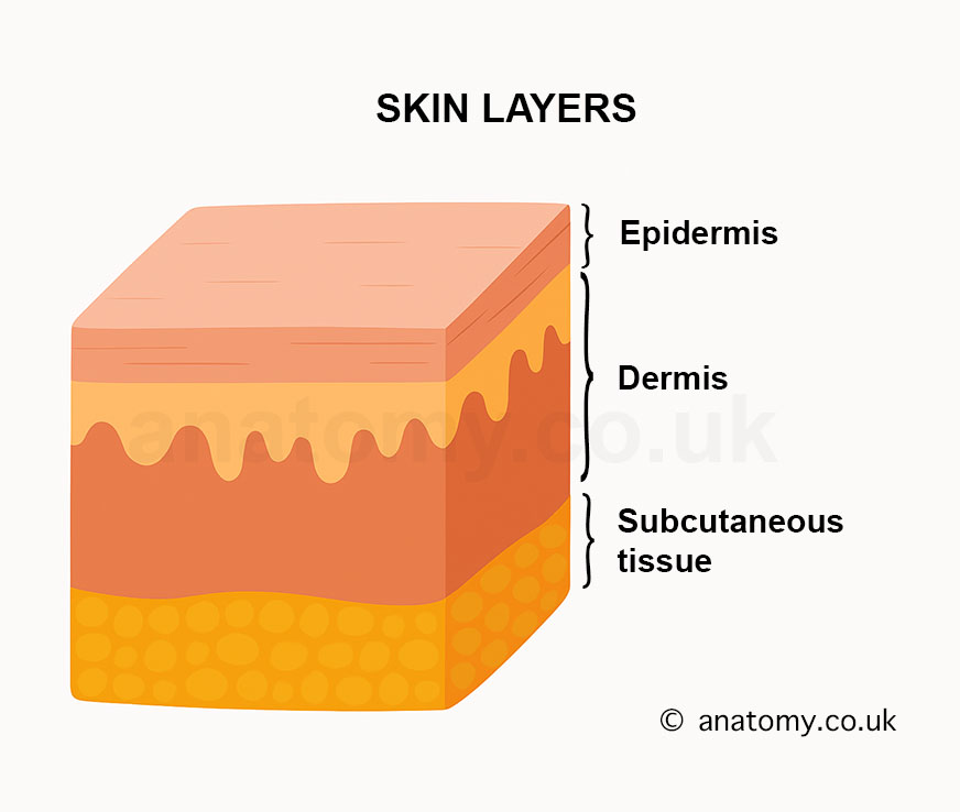
Skin is the largest organ of the human body, serving as a protective covering that encases and safeguards internal structures. It is composed of three primary layers: the epidermis (outer layer), the dermis (middle layer), and the subcutaneous tissue (innermost layer). These layers work together to maintain the body's integrity, protect against environmental threats, regulate temperature, and facilitate sensory perception. The skin varies in thickness across the body, being thicker on areas like the palms and soles, and thinner on areas like the eyelids.
Location
Skin covers the entire external surface of the body. It forms a continuous protective layer from the top of the head to the soles of the feet, including all external body parts. Skin thickness and structure vary depending on location and the function of the specific body area.
Structure and Anatomy
The skin is a highly complex organ that serves as the body's protective outer layer. It consists of multiple layers and specialized structures that support its various roles. Below is a detailed breakdown of the anatomy of the skin:
Layers of the Skin
The skin is composed of three main layers: the epidermis, the dermis, and the subcutaneous tissue (also known as the hypodermis). Each layer has a distinct structure and specific components that contribute to the overall function of the skin.
Epidermis
Outermost Layer: The epidermis is the topmost layer of the skin, responsible for creating a protective barrier. It is made up of keratinized stratified squamous epithelium, meaning it is composed of multiple layers of flat cells that are filled with keratin, a tough, fibrous protein.
Cell Layers: The epidermis consists of several sublayers:
Stratum Corneum: The outermost layer of dead, flattened cells (corneocytes) that are constantly shed and replaced.
Stratum Lucidum: A clear, thin layer found only in thick skin, such as the palms and soles.
Stratum Granulosum: Cells in this layer contain keratohyalin granules and begin to die as they move upwards.
Stratum Spinosum: This layer contains keratinocytes connected by desmosomes, giving them a spiny appearance.
Stratum Basale: The deepest layer, where new keratinocytes are produced. It also contains melanocytes, which produce melanin, and Merkel cells, which function in touch sensation.
Key Cells: The epidermis contains keratinocytes, melanocytes, Langerhans cells (immune cells), and Merkel cells (sensory cells).
Dermis
Middle Layer: The dermis lies beneath the epidermis and is much thicker. It provides strength and elasticity to the skin, thanks to the collagen and elastin fibers present in this layer.
Papillary Dermis: The upper layer of the dermis, characterized by dermal papillae, which form ridges that interlock with the epidermis. This layer contains capillaries that nourish the epidermis, and Meissner’s corpuscles, responsible for sensing light touch.
Reticular Dermis: The deeper portion of the dermis, consisting of dense irregular connective tissue. It contains thicker collagen fibers, elastin fibers, and other skin appendages such as hair follicles, sebaceous (oil) glands, and sweat glands. Pacinian corpuscles found in this layer detect deep pressure and vibrations.
Blood Supply and Nerves: The dermis is rich in blood vessels that supply nutrients to both the dermis and the overlying avascular epidermis. It also houses an extensive network of nerve endings responsible for sensation, including pain, temperature, and tactile stimuli.
Subcutaneous Tissue (Hypodermis)
Deepest Layer: The subcutaneous tissue, also called the hypodermis, lies beneath the dermis and is primarily composed of adipose tissue (fat cells), connective tissue, blood vessels, and nerves. This layer acts as an energy reserve, provides insulation, and cushions underlying structures such as muscles and bones.
Fat Storage: The adipose tissue in this layer serves as a site for fat storage, which provides energy and helps regulate body temperature by insulating the body. It also serves as a shock absorber, protecting internal organs from mechanical injuries.
Blood Supply: Larger blood vessels run through the subcutaneous tissue, branching off into smaller vessels that supply the dermis and epidermis.
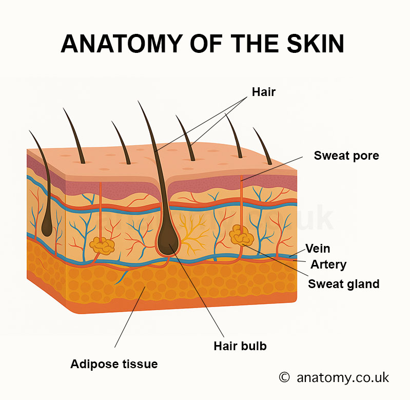
Skin Appendages
The skin contains several appendages that are derived from the epidermis and extend into the dermis. These include:
Hair Follicles
Hair Growth: Hair follicles are tubular structures located in the dermis and subcutaneous tissue, responsible for producing hair. Each follicle is associated with a sebaceous gland and a small muscle called the arrector pili.
Structure: The hair follicle consists of the hair bulb, where the hair matrix cells divide and grow, and the dermal papilla, which supplies nutrients to the growing hair. Hair emerges from the follicle through the hair shaft.
Associated Structures: Arrector pili muscles are attached to hair follicles and can contract to make the hair stand up (a reaction known as piloerection, or goosebumps).
Sebaceous Glands
Oil Production: Sebaceous glands are located in the dermis and are associated with hair follicles.[7] These glands produce sebum, an oily substance that helps lubricate and waterproof the skin and hair.
Holocrine Secretion: Sebaceous glands release sebum through holocrine secretion, a process in which cells in the gland break down and release their contents into the hair follicle.
Sweat Glands
Eccrine Sweat Glands: Eccrine glands are widely distributed across the skin and are responsible for thermoregulation through the production of sweat. These glands secrete a watery fluid directly onto the skin surface via sweat ducts.
Apocrine Sweat Glands: Apocrine glands are found in specific areas, such as the armpits and groin. They produce a thicker, more viscous sweat that is released into hair follicles and can contribute to body odor when broken down by bacteria.
Blood and Lymphatic Vessels
Blood Supply: The skin contains a network of blood vessels that extend from the subcutaneous tissue into the dermis. These vessels provide essential nutrients and oxygen to the skin and help regulate body temperature through vasoconstriction (narrowing) and vasodilation (widening) of the blood vessels.
Lymphatic Vessels: The skin is also equipped with lymphatic vessels, which help drain excess fluid and remove waste products from the skin, aiding in immune responses and maintaining tissue health.
Nerve Endings and Sensory Receptors
Sensory Perception: The skin contains a variety of sensory receptors that allow it to detect stimuli such as touch, pressure, pain, and temperature. These receptors include:
Meissner's Corpuscles: Located in the papillary dermis, these receptors detect light touch.
Pacinian Corpuscles: Found in the deeper dermis and subcutaneous layer, they respond to deep pressure and vibration.[6]
Free Nerve Endings: These receptors detect pain, temperature, and certain touch stimuli and are distributed throughout the dermis and epidermis.
Pigmentation
Melanin Production: Melanocytes, located in the stratum basale of the epidermis, produce melanin, the pigment responsible for skin color. Melanin protects the skin from ultraviolet (UV) radiation by absorbing and dispersing harmful rays.
Skin Tone Variation: Differences in skin tone among individuals are due to variations in the amount, type, and distribution of melanin produced by melanocytes.
Basement Membrane
Epidermis-Dermis Interface: The basement membrane is a thin layer that separates the epidermis from the dermis. It is composed of specialized proteins, including collagen, laminin, and proteoglycans, which provide structural support and anchor the epidermis to the dermis.
Cell Adhesion: Hemidesmosomes in the basal keratinocytes of the epidermis and anchoring fibrils in the dermis attach the two layers, ensuring that they remain connected during movement and mechanical stress.[8]
Skin Variability Across the Body
Thick vs. Thin Skin: The thickness of the skin varies depending on its location. Thick skin is found on the palms of the hands and soles of the feet, and it has a thicker epidermis with a prominent stratum lucidum. Thin skin, found on the rest of the body, has a thinner epidermis and lacks the stratum lucidum.
Variation in Gland Density: The distribution and density of sebaceous and sweat glands vary across the body. For example, the face and scalp have a high density of sebaceous glands, while the palms and soles have a high concentration of eccrine sweat glands but no sebaceous glands.
Function
The skin is the body’s largest organ, and it performs a wide variety of functions that are essential for maintaining overall health and well-being. These functions range from protecting the body to regulating temperature and enabling sensory perception. Below is a detailed breakdown of the skin’s functions:
Protection
Physical Barrier: The skin serves as the body’s primary protective barrier against mechanical damage, pathogens, and harmful substances. The outermost layer of the skin, the epidermis, consists of keratinized cells that form a tough, waterproof shield. This prevents harmful microorganisms, chemicals, and physical trauma from penetrating deeper tissues.
Chemical Protection: The acid mantle, a slightly acidic layer formed by sweat and sebum on the skin’s surface, inhibits the growth of harmful bacteria and fungi. The acidic environment helps maintain a healthy skin microbiome while preventing infections.[5]
UV Radiation Protection: Melanocytes, located in the stratum basale of the epidermis, produce melanin, a pigment that absorbs and scatters harmful ultraviolet (UV) radiation. Melanin protects the skin from UV-induced DNA damage, which can lead to skin cancer and premature aging.
Sensation and Sensory Perception
Touch and Pressure: The skin is equipped with numerous sensory receptors that detect tactile stimuli. These receptors include Meissner's corpuscles in the dermis, which respond to light touch, and Pacinian corpuscles, which detect deep pressure and vibrations. This allows the skin to sense textures, pressure, and movement.
Temperature Sensation: Thermoreceptors in the skin detect changes in temperature, enabling the body to sense heat and cold. These receptors help maintain thermal homeostasis by triggering appropriate responses, such as sweating or shivering, when environmental conditions change.
Pain and Injury Detection: The skin contains nociceptors, specialized nerve endings that detect painful stimuli such as cuts, burns, or extreme temperatures. These receptors send signals to the brain, initiating protective responses that prevent further damage to the body.
Thermoregulation
Sweat Production and Heat Loss: The skin plays a central role in temperature regulation through the production of sweat by eccrine sweat glands. When the body becomes overheated, sweat is produced and released onto the skin's surface. As sweat evaporates, it cools the body, helping to maintain a stable internal temperature.
Vasodilation and Vasoconstriction: Blood vessels in the dermis help regulate body temperature by undergoing vasodilation (widening) and vasoconstriction (narrowing). When the body is hot, blood vessels dilate, increasing blood flow to the skin’s surface, where heat is dissipated. In contrast, when the body is cold, blood vessels constrict to reduce heat loss by minimizing blood flow to the skin.
Piloerection (Goosebumps): Arrector pili muscles attached to hair follicles contract in response to cold or emotional stimuli, causing the hair to stand upright (goosebumps). In humans, this response has a minimal effect, but in animals, it helps trap heat by creating an insulating layer of air near the skin.[4]
Water Retention and Hydration
Preventing Water Loss: The skin acts as a waterproof barrier that prevents excessive loss of water from the body. The stratum corneum, the outermost layer of the epidermis, contains lipids and keratinized cells that create a hydrophobic barrier, limiting transepidermal water loss. This helps maintain proper hydration levels and prevents dehydration.
Hydration Balance: Sebum, produced by sebaceous glands, and the lipids secreted by keratinocytes help retain moisture in the skin. This balance of oil and water ensures that the skin remains supple and does not become excessively dry or prone to cracking.
Vitamin D Synthesis
UV-Induced Vitamin D Production: The skin plays a critical role in the synthesis of vitamin D, which is essential for calcium absorption and bone health. When exposed to UVB radiation from sunlight, the keratinocytes in the epidermis convert 7-dehydrocholesterol into vitamin D3 (cholecalciferol). This vitamin is then processed by the liver and kidneys to produce its active form, calcitriol, which regulates calcium and phosphate levels in the body.
Immune Defense
Langerhans Cells: The skin contains specialized immune cells called Langerhans cells, which reside in the stratum spinosum of the epidermis. These cells capture and process antigens (foreign substances) that enter the skin, presenting them to T cells to initiate an immune response. This makes the skin an important part of the immune system, providing a first line of defense against infections.
Barrier Against Pathogens: The tight junctions between keratinocytes, along with the presence of antimicrobial peptides in the skin’s surface, help prevent the entry of pathogens such as bacteria, viruses, and fungi.[3] The skin’s role as a barrier is crucial for preventing infections and maintaining overall health.
Excretion and Waste Removal
Sweat Gland Function: The skin aids in the excretion of waste products through eccrine sweat glands. These glands secrete sweat that contains water, salts, urea, and other metabolic waste products. This helps in the removal of small amounts of toxins from the body and maintains electrolyte balance.
Regulation of Salt Levels: In addition to excreting waste, sweat helps regulate sodium chloride (salt) levels in the body. By adjusting the concentration of salt in sweat, the skin assists in maintaining proper electrolyte balance, which is essential for normal physiological function.
Absorption
Transdermal Absorption: While the skin primarily functions as a barrier, it can also absorb certain substances. Lipid-soluble substances, such as certain medications, chemicals, and vitamins, can pass through the skin layers and enter the bloodstream. This principle is used in transdermal drug delivery systems, such as nicotine patches or hormone replacement therapy patches.
Limited Absorption of Water-Soluble Substances: While the skin is largely impermeable to water-soluble substances, certain small molecules can be absorbed under specific conditions, such as when the skin is broken or in the presence of enhancers that increase skin permeability.
Fat Storage and Cushioning
Subcutaneous Fat Storage: The subcutaneous tissue (hypodermis), the deepest layer of the skin, contains adipose tissue, which stores fat. This fat serves as an energy reserve, providing a source of stored energy that can be used when needed by the body. It also insulates the body against cold temperatures.
Cushioning and Shock Absorption: The fat in the subcutaneous layer provides cushioning that helps protect internal organs, bones, and muscles from mechanical shocks or impacts.[2] This is especially important in areas of the body that are prone to pressure or trauma, such as the buttocks and heels.
Aesthetic and Social Functions
Expression of Identity: The skin plays a significant role in appearance and personal identity. Variations in skin color, texture, and hair growth contribute to individual differences in physical appearance. Skin color is determined by the amount and type of melanin produced by melanocytes, while the texture of the skin can be influenced by factors such as hydration and oil production.
Emotional Expression: The skin is involved in the display of emotions. Blushing, caused by the dilation of blood vessels in the skin, occurs in response to emotions like embarrassment or excitement. Goosebumps, caused by the contraction of arrector pili muscles, can also be triggered by emotional stimuli such as fear or awe.
Regulation of Blood Flow
Vascular Regulation: The blood vessels in the dermis play a critical role in regulating blood flow. In response to heat or cold, the skin’s blood vessels either dilate or constrict to control blood flow and regulate body temperature.[1] This also helps manage blood pressure and ensures that organs and tissues receive the appropriate amount of blood under varying conditions.
Wound Healing and Tissue Repair
Wound Healing Process: When the skin is damaged, the epidermis and dermis work together to heal wounds and regenerate new tissue. Fibroblasts in the dermis produce collagen and elastin, which help repair the skin and form scar tissue. Keratinocytes migrate to the wound site, covering it and restoring the protective barrier.
Scar Formation: If the dermis is damaged, scar tissue may form. This tissue, which is rich in collagen but lacks the full structure and function of normal skin, helps close the wound but may result in altered texture and appearance.
Hair and Nail Growth
Hair and Nail Protection: The skin supports the growth of hair and nails, both of which serve protective functions. Hair provides insulation and shields sensitive areas like the scalp, while nails protect the fingertips and toes from injury and enhance tactile sensation.
Clinical Significance
The skin's role as the body's largest organ makes it crucial in various clinical contexts. Disorders affecting the skin can range from mild, like acne or dermatitis, to life-threatening conditions such as melanoma or burns. The skin serves as an indicator of overall health, with conditions like jaundice, cyanosis, and pallor often signaling systemic diseases such as liver dysfunction, oxygen deficiency, or anemia. Skin integrity is vital for preventing infections, as damage to the skin barrier—through wounds, ulcers, or burns—can lead to complications like sepsis. Chronic conditions such as eczema, psoriasis, and rosacea impact quality of life and require long-term management. Skin cancer, particularly melanoma, is a major concern due to prolonged UV exposure.
Published on November 29, 2024
Last updated on May 17, 2025
Last updated on May 17, 2025