CC
Cricoid Cartilage
Ring-shaped cartilage of larynx supporting airway
RegionHead and Neck
SystemRespiratory System
The cricoid cartilage is a critical structure located in the larynx or voice box. Distinguished by its ring-like shape, it is the only complete cartilaginous ring that encircles the airway. This cartilage provides foundational support to the larynx and plays a vital role in breathing and speech functions. Located inferior to the thyroid cartilage and superior to the trachea, the cricoid cartilage forms the base of the laryngeal cartilages and is essential for maintaining airway patency. Additionally, its articulation with other laryngeal cartilages and its function as an attachment point for several muscles emphasize its significance in the anatomy of the neck.
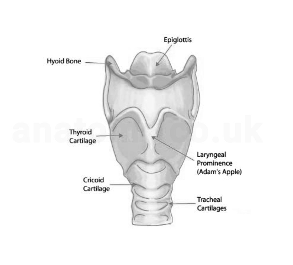
Image 1: Laryngeal cartilages overview
Anatomy of the Cricoid Cartilage
The cricoid cartilage is a distinct structure within the larynx due to its complete ring-like formation. Understanding its specific anatomical features is essential for a comprehensive grasp of laryngeal functions and clinical interventions.
Image 2: Cricoid cartilage diagram
Shape and Structure
The cricoid cartilage is shaped like a signet ring. Its broad posterior section, known as the lamina, provides a vertical plate-like structure. The narrower anterior part, referred to as the arch, offers the band-like aspect of the ring.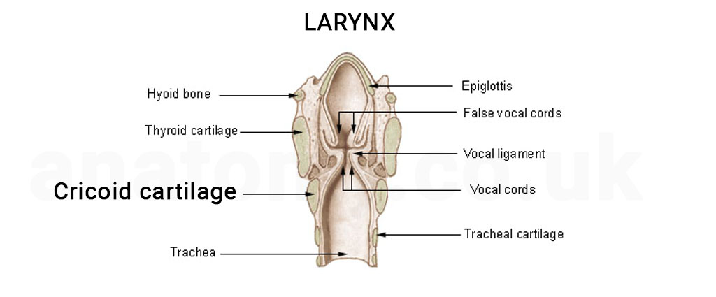
Image 3: Detailed cricoid cartilage anatomy Diagram
Location
The cricoid cartilage is positioned below the thyroid cartilage and above the trachea's first ring, making it the lowermost component of the laryngeal cartilages.Articulations
On its lateral sides, the cricoid cartilage features articular facets that allow it to connect with the arytenoid cartilages. These articulations are essential for the movement and function of the vocal cords.Dimensions
The lamina (posterior part) is typically broader in height than the arch (anterior part). In adults, the height of the lamina averages around 2-3cm, while the arch is approximately 1cm in height.Attachments
Several intrinsic laryngeal muscles, like the posterior cricoarytenoid and lateral cricoarytenoid, attach to the cricoid cartilage. The cricothyroid ligament connects it to the thyroid cartilage above, and the cricotracheal ligament attaches it to the trachea below.Development and Variation
Gaining insights into the developmental stages and variations of the cricoid cartilage provides a foundational understanding of its formation and potential anatomical differences.Embryological Development
The cricoid cartilage begins to form during the embryonic stage. It originates from the fourth branchial arch's mesenchymal tissue. As embryonic development progresses, this tissue condenses and undergoes chondrification, leading to the formation of the cricoid cartilage.Maturation
The cricoid cartilage continues to grow and mature through childhood. By late adolescence, it typically reaches its full size and shape, ready to perform its pivotal role in the adult larynx.Anatomical Variations
While the cricoid cartilage generally maintains a consistent ring-like structure, minor anatomical variations can occur. These might include slight differences in thickness or subtle asymmetries in the lamina or arch. However, these variations are typically benign and do not affect function.Age-related Changes
Similar to other cartilages in the body, the cricoid cartilage can undergo age-associated changes. Over time, parts of the cartilage might exhibit calcification, leading to increased rigidity. This process can be more pronounced in older adults.Functions
The cricoid cartilage, given its strategic position and unique ring-like structure, has a myriad of functions crucial for respiratory and vocal activities.Structural Support
Serving as the only complete ring in the larynx, the cricoid cartilage offers structural integrity to the larynx. It forms the base upon which other laryngeal cartilages rest and articulate.Protection
Being a sturdy cartilaginous ring, the cricoid cartilage acts as a protective barrier, safeguarding the airway and underlying structures from external traumas or compressive forces.Vocal Cord Articulation
The cricoid cartilage provides attachment points for the arytenoid cartilages on its upper lateral surfaces. These arytenoid cartilages, in turn, anchor the vocal cords, allowing for their movement and thus playing an essential role in modulating voice.Muscle Attachment
Several intrinsic laryngeal muscles, such as the cricothyroid and the posterior cricoarytenoid, attach to the cricoid cartilage. These muscles facilitate various movements of the larynx, including adjusting tension on the vocal cords and opening or closing the glottis.Clinical Relevance
The cricoid cartilage, due to its unique position and structure, has multiple clinical implications, making it an essential focus in various medical situations and procedures.Cricoid Pressure (Sellick's Maneuver)
In anesthesiology, the application of pressure to the cricoid cartilage, known as Sellick's maneuver, is sometimes used during rapid sequence intubation to prevent regurgitation and aspiration of stomach contents. The pressure compresses the esophagus against the vertebral column, serving as a preventive measure.[8]Cricothyrotomy
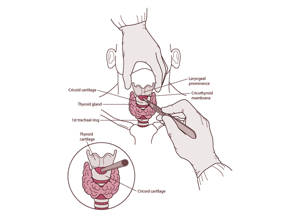
Image 4: Cricothyrotomy procedure
In emergency situations where securing an airway is challenging using conventional methods, a cricothyrotomy can be performed.[6] This procedure involves making an incision through the cricothyroid membrane, which lies between the cricoid and thyroid cartilages, to establish an emergency airway.Injuries and Fractures
Due to its anterior position, the cricoid cartilage can be susceptible to traumatic injuries, especially in cases of blunt trauma to the neck.[4] Fractures or disruptions of the cricoid can lead to airway compromise and require urgent medical intervention.Laryngeal Surgeries
The cricoid cartilage often plays a central role in surgeries related to the larynx, such as in cases of laryngeal tumors or reconstructive procedures. Understanding its anatomy and relations is pivotal for surgeons working in the region.Diagnostic Imaging
In radiological studies, the cricoid cartilage serves as an easily identifiable landmark, helping in the evaluation of neck and laryngeal pathologies. Calcification or anomalies of the cricoid cartilage can be visualized using techniques like X-rays, CT scans, or MRIs.[1]Interesting Facts and Myths
The cricoid cartilage, while primarily known for its clinical and anatomical significance, has also been surrounded by various facts and myths. Name Origin: The term "cricoid" is derived from the Greek word "krikoeides," which means "ring-like." This name aptly describes its unique ring-shaped structure among the laryngeal cartilages.[7] Sole Complete Ring: The cricoid cartilage stands out as the only cartilage in the airway to form a complete ring. This distinctive shape makes it pivotal in maintaining the patency of the airway. Cricoid Pressure Controversies: While the Sellick's maneuver (cricoid pressure) is a widely recognized technique, especially in anesthesiology, its efficacy and safety have been subjects of debate. Some studies suggest potential risks, including esophageal rupture, while others emphasize its utility in preventing aspiration. Age-old Recognition: Historical medical texts, dating back centuries, have referenced the cricoid cartilage, indicating its recognition and importance in medical history.[3] Myth: Cricoid Cartilage Prevents Choking: A common misconception is that the cricoid cartilage acts as a protective barrier against choking.[5] While it plays a role in maintaining airway structure, the act of swallowing and preventing aspiration primarily involves physiological reflexes and other anatomical structures, rather than the cricoid cartilage itself.Frequently Asked Questions
Where is the cricoid cartilage located in relation to other laryngeal structures?
The cricoid cartilage is positioned below the thyroid cartilage and above the trachea's first ring. It forms the base of the laryngeal cartilages.[2]Why is the cricoid cartilage often emphasized in emergency airway procedures?
Answer: Due to its position and ring-like structure, the cricoid cartilage serves as a landmark in emergency airway procedures like cricothyrotomy. Its unique shape and location make it identifiable, aiding in the accurate establishment of an emergency airway.What is the significance of the cricoid cartilage's ring-like structure?
The cricoid cartilage's ring-like structure ensures that the airway remains open and patent. As the only complete cartilaginous ring in the airway, it offers structural integrity and protection to the larynx.Are there common diseases or conditions that specifically affect the cricoid cartilage?
While the cricoid cartilage can be affected by general laryngeal conditions like tumors, inflammations, or traumatic injuries, it's not commonly associated with specific diseases. However, its involvement in laryngeal surgeries or trauma can lead to conditions like subglottic stenosis.How does the cricoid cartilage differ from the thyroid cartilage?
The most notable difference is their shape. The cricoid cartilage is ring-like and completely encircles the airway, while the thyroid cartilage is shield-shaped and does not form a complete ring. The cricoid is located inferior to the thyroid and forms the base of the laryngeal cartilages.Published on October 9, 2023
Last updated on May 11, 2025
Last updated on May 11, 2025