L
Lung
Primary organ for respiration located in thorax
RegionThorax
SystemRespiratory System
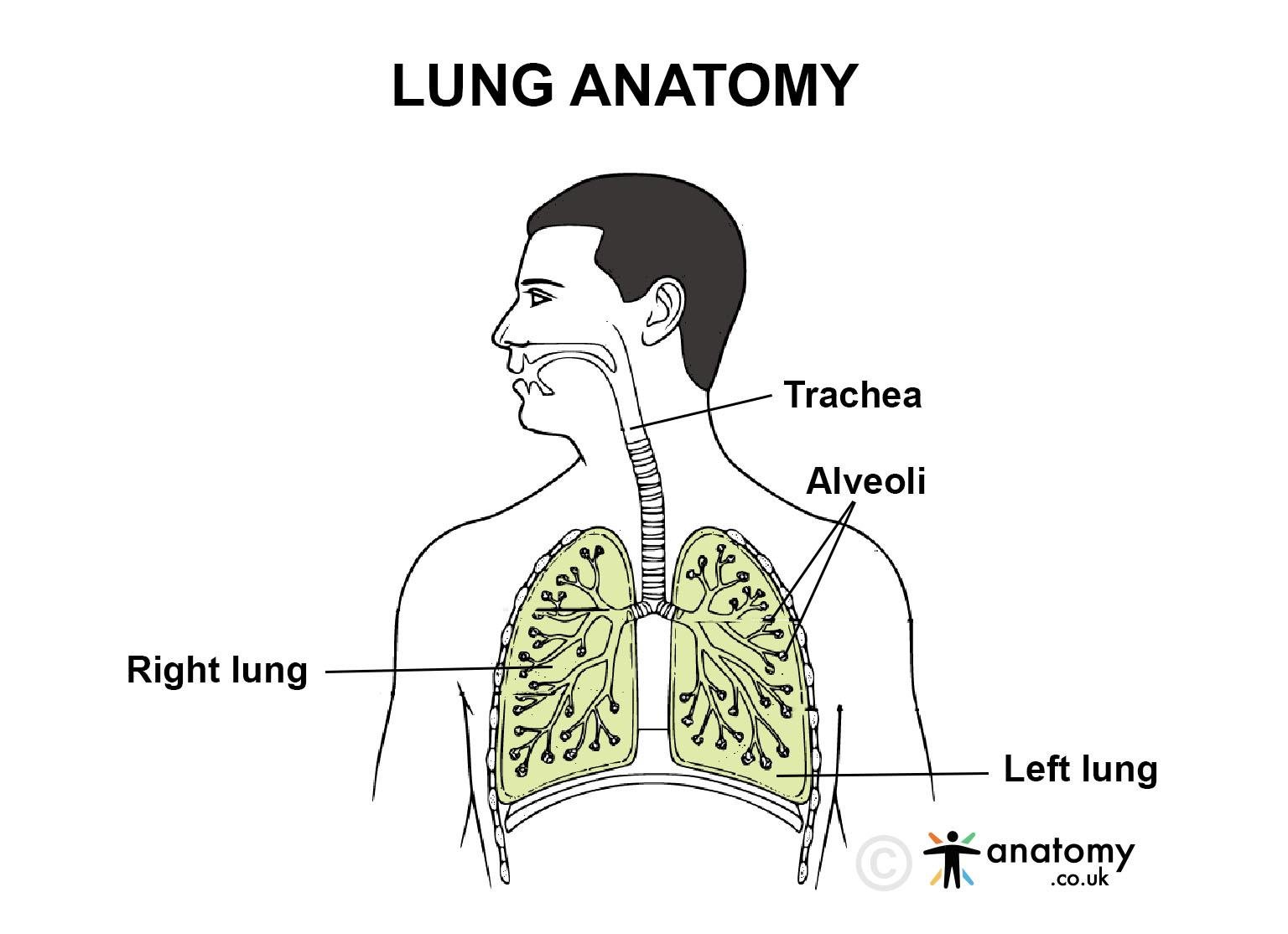
The lung is a vital organ in the respiratory system responsible for the exchange of oxygen and carbon dioxide between the air we breathe and the bloodstream.[2] Humans have two lungs, which are soft, spongy, and cone-shaped. Each lung is divided into lobes: the right lung has three lobes, while the left lung has two lobes, making room for the heart.


Location
The lungs are located in the thoracic cavity, on either side of the heart. They are enclosed by the rib cage and rest on the diaphragm, a muscle that aids in breathing. The lungs are protected by a thin membrane called the pleura.Anatomy
The lungs are highly specialized organs with a complex structure designed for efficient gas exchange. Below is a detailed exploration of their anatomy.Gross Anatomy
- Lobes: The lungs are divided into lobes. The right lung has three lobes—superior, middle, and inferior—while the left lung has two lobes—superior and inferior. The left lung is slightly smaller than the right lung to make space for the heart.
- Fissures: The lobes of the lungs are separated by fissures. In the right lung, the horizontal fissure divides the superior and middle lobes, and the oblique fissure separates the middle and inferior lobes.[3] In the left lung, only the oblique fissure separates the superior and inferior lobes.
Pleura
- Visceral Pleura: The outer surface of the lungs is covered by a thin, slippery membrane called the visceral pleura. This membrane adheres to the lung tissue and helps reduce friction during breathing.
- Parietal Pleura: The parietal pleura lines the inner surface of the thoracic cavity, the diaphragm, and the mediastinum. Between the visceral and parietal pleura is the pleural cavity, filled with pleural fluid that acts as a lubricant and ensures smooth movement of the lungs during breathing.
Bronchial Tree
The lungs receive air through a branching system of airways called the bronchial tree, which starts from the trachea and divides into smaller and smaller tubes.- Primary Bronchi: The trachea divides into two primary bronchi (left and right), one for each lung. The right bronchus is shorter, wider, and more vertical than the left.
- Secondary (Lobar) Bronchi: Each primary bronchus branches into secondary bronchi, which correspond to the lobes of the lungs. The right lung has three secondary bronchi, and the left lung has two.
- Tertiary (Segmental) Bronchi: The secondary bronchi further divide into tertiary bronchi, which supply air to specific segments within each lobe, known as bronchopulmonary segments.
- Bronchioles: The tertiary bronchi further divide into bronchioles, which are smaller airways that lack cartilage.[1] These bronchioles continue to branch and eventually lead to terminal bronchioles, which mark the end of the conducting airways.
- Respiratory Bronchioles: These lead from the terminal bronchioles to the alveolar ducts and alveolar sacs, where gas exchange occurs.
Alveoli
- Alveolar Sacs: The alveoli are tiny air sacs located at the ends of the respiratory bronchioles. Alveoli are grouped together into clusters called alveolar sacs. Each alveolus is surrounded by a network of capillaries, facilitating gas exchange between the air and blood.[4]
- Alveolar Wall: The alveolar wall is made of a thin layer of simple squamous epithelium. This thin barrier allows for efficient diffusion of gases. The alveoli also contain surfactant-producing cells that help reduce surface tension, preventing alveolar collapse during exhalation.
Blood Supply
The lungs receive blood through two circulatory systems:- Pulmonary Circulation: Deoxygenated blood from the heart is pumped into the lungs via the pulmonary arteries. This blood travels through the capillaries surrounding the alveoli, where gas exchange occurs, and oxygenated blood returns to the heart via the pulmonary veins.
- Bronchial Circulation: The lung tissue itself is nourished by the bronchial arteries, which arise from the aorta. The bronchial veins carry deoxygenated blood away from the lung tissue and drain into the azygos venous system.
Lymphatic System
The lungs are equipped with an extensive lymphatic system to remove excess fluid and filter foreign particles. Superficial lymphatic vessels drain the outer surface of the lungs, while deep lymphatic vessels drain the lung parenchyma and surrounding connective tissue. The lymph is transported to the hilar lymph nodes at the root of the lung, and eventually into the thoracic duct or right lymphatic duct.Nervous Innervation
The lungs are innervated by both the sympathetic and parasympathetic nervous systems.- Sympathetic Innervation: Sympathetic nerve fibers cause bronchodilation, which increases airflow by relaxing the smooth muscles in the bronchioles.
- Parasympathetic Innervation: Parasympathetic fibers from the vagus nerve stimulate bronchoconstriction, which reduces airflow by contracting the smooth muscles of the bronchioles. This innervation also controls mucus secretion in the airways.
Lung Segments
Each lung lobe is further divided into functional units called bronchopulmonary segments, each supplied by its own tertiary bronchus and artery. These segments are important in clinical settings, as they can be surgically removed if damaged without affecting neighboring segments.- The right lung has 10 bronchopulmonary segments: 3 in the superior lobe, 2 in the middle lobe, and 5 in the inferior lobe.
- The left lung typically has 8 to 10 segments: 4 to 5 in the superior lobe and 4 to 5 in the inferior lobe.
Function
The lungs are vital organs responsible for several key functions that are crucial to maintaining life. Below is a detailed explanation of the various functions of the lungs.Gas Exchange
The primary function of the lungs is to facilitate the exchange of gases, specifically oxygen and carbon dioxide, between the air we breathe and the bloodstream.- Oxygen Intake: During inhalation, oxygen-rich air enters the lungs and travels down through the bronchial tree to the alveoli.[5] Oxygen from the air diffuses through the thin walls of the alveoli and into the surrounding capillaries. It binds to hemoglobin in red blood cells and is then transported throughout the body to tissues and organs.
- Carbon Dioxide Removal: Simultaneously, carbon dioxide, a waste product of cellular metabolism, is carried in the blood from the body’s tissues to the lungs. In the alveoli, carbon dioxide diffuses from the capillaries into the alveolar air and is expelled during exhalation.
Regulation of Blood pH
The lungs play a critical role in maintaining the acid-base balance (pH) of the blood. This is achieved by regulating the levels of carbon dioxide, which is acidic when dissolved in blood.- CO₂ Removal: When carbon dioxide levels in the blood rise, the pH decreases (becomes more acidic). The lungs respond by increasing the rate and depth of breathing (hyperventilation) to expel more carbon dioxide, thus raising the pH back to normal.
- Breath Control: Conversely, if carbon dioxide levels drop too low (causing an increase in pH, or alkalosis), the lungs slow down breathing (hypoventilation), allowing carbon dioxide to accumulate and the pH to return to normal levels.
Protection Against Inhaled Particles and Pathogens
The lungs serve as a defense system against harmful particles, pathogens, and irritants that enter the respiratory system through inhalation.- Mucociliary Clearance: The airways are lined with mucus-producing cells and ciliated epithelium. The mucus traps dust, microbes, and other foreign particles, while the cilia (tiny hair-like structures) move the mucus upwards toward the throat, where it can be swallowed or coughed out.
- Immune Defense: The lungs contain immune cells, such as alveolar macrophages, which patrol the alveoli and engulf harmful microorganisms, particles, and debris. These cells are the first line of defense against respiratory infections and pollutants.
Filtration of Blood Clots and Small Emboli
The lungs act as a filter for the blood, helping to trap small clots or air bubbles (emboli) that may have entered the bloodstream.[6] Pulmonary Capillary Filter: As blood passes through the pulmonary capillaries, the lungs can trap and break down small blood clots or foreign particles before they reach critical areas of the body, such as the brain or heart.Metabolism of Bioactive Substances
The lungs participate in the metabolism and modification of various substances circulating in the blood. They produce, store, and modify several bioactive compounds.- Angiotensin-Converting Enzyme (ACE): One of the best-known examples is the production of angiotensin-converting enzyme (ACE) in the pulmonary capillaries. ACE converts angiotensin I into angiotensin II, a potent vasoconstrictor that helps regulate blood pressure.
- Inactivation of Substances: The lungs can also inactivate or remove certain substances from the blood, such as bradykinin, serotonin, and prostaglandins, which affect blood vessel tone and other physiological processes.
Maintenance of Temperature and Humidity of Air
As air passes through the nasal passages and into the lungs, it is warmed and humidified to match body temperature and moisture levels, ensuring that the alveolar surfaces and delicate lung tissues are not irritated or damaged by dry or cold air.- Humidification: The moist mucous membranes lining the respiratory tract add water vapor to the air we inhale, preventing the drying out of the lungs and maintaining a comfortable environment for gas exchange.
- Warming of Air: The blood vessels in the nasal passages and bronchi warm the air to body temperature, ensuring that the air reaching the alveoli is at an optimal temperature for efficient gas exchange.
Vocalization (Phonation)
The lungs play an indirect but crucial role in speech and vocalization.- Airflow for Sound Production: Air expelled from the lungs flows through the larynx (voice box), where it passes over the vocal cords, causing them to vibrate. These vibrations produce sound, and the modulation of airflow from the lungs allows for different tones and pitches in speech or singing.
- Control of Exhalation: The lungs regulate the force of air exhaled, controlling the volume and intensity of sound produced. This controlled exhalation is essential for clear vocalization.
Olfaction (Sense of Smell)
Although not directly part of the lung function, inhalation through the nose allows air to pass over the olfactory receptors in the nasal cavity, facilitating the sense of smell. This process is integral to respiratory function, as it helps detect harmful substances and enhances the overall sensory experience.Maintenance of Blood Volume and Pressure
The lungs indirectly influence blood pressure and volume through the regulation of substances such as angiotensin II and the balance of oxygen and carbon dioxide levels. This contributes to overall cardiovascular health.[8] Influence on Blood Volume: By regulating the amount of blood flow to the lungs and their capillary networks, the lungs can adjust blood volume levels that affect overall blood pressure and circulation.Clinical Significance
The lungs are essential for life, and their dysfunction can lead to a wide range of respiratory and systemic diseases. Conditions like chronic obstructive pulmonary disease (COPD), asthma, and lung cancer are common and can severely impact lung function. Chronic bronchitis and emphysema, both under the umbrella of COPD, cause obstruction of airflow, making breathing difficult. Pneumonia, an infection of the lung tissue, can cause severe inflammation, while pulmonary fibrosis leads to scarring of lung tissue, reducing its ability to expand and contract.[7] Lung diseases can also lead to complications such as respiratory failure, where the lungs fail to adequately oxygenate the blood, and pulmonary hypertension, an increase in blood pressure within the pulmonary arteries. Early diagnosis and treatment of lung-related conditions are critical to preventing irreversible damage and maintaining overall health. Regular screening, such as spirometry and chest X-rays, can help in the early detection of these conditions.Published on November 29, 2024
Last updated on May 17, 2025
Last updated on May 17, 2025