CJ
Cricothyroid Joint
Joint between cricoid and thyroid cartilages
RegionHead and Neck
SystemMusculoskeletal System
The cricothyroid joint is a pivotal articulation within the larynx, responsible for facilitating a range of movements that play a vital role in voice modulation. Situated between the cricoid cartilage and the thyroid cartilage, this diarthrodial, synovial joint allows for pivotal and gliding motions that modulate tension on the vocal cords.
Comprising of two articular surfaces, the joint is formed by the articulation between the cricoid cartilage's lateral aspect and the inferior cornu and lower lamina of the thyroid cartilage. Capsular ligaments, which are thicker anteriorly than posteriorly, envelop the joint and provide stability.
The cricothyroid joint's ability to tilt the thyroid cartilage anteriorly and inferiorly relative to the cricoid is crucial in adjusting the vocal cord's tension. This dynamic function is fundamental in varying pitch during phonation.
This joint, while small and seemingly simple, has a profound influence on the complexities of voice production, emphasizing its importance in both anatomy and clinical practice.
Anatomy

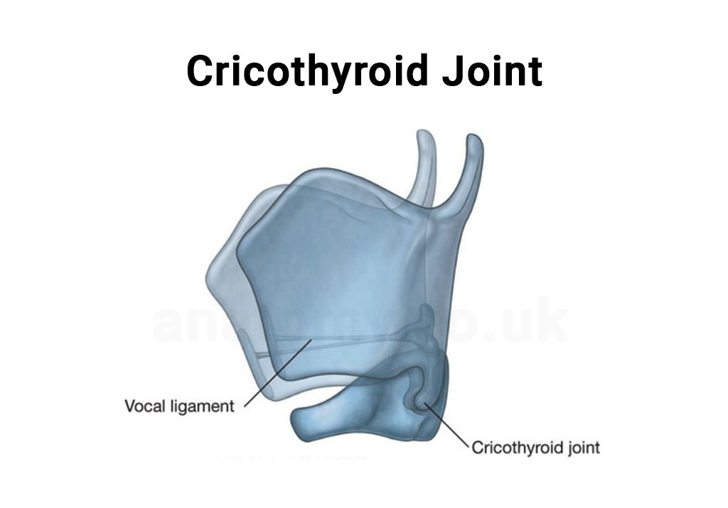
Image 1: Location of Cricothyroid Joint
Located within the intricate framework of the larynx, the cricothyroid joint occupies a strategic position that enables its essential function in voice modulation.Relation to Laryngeal Cartilages
The joint is formed at the point where the cricoid and thyroid cartilages meet. Specifically, it's the articulation between the lateral aspect of the cricoid cartilage and the inferior cornu and lower lamina of the thyroid cartilage.Vertical Position
In the vertical plane, the cricothyroid joint sits above the trachea and below the broader structure of the larynx. It's positioned superiorly to the first tracheal ring and inferiorly to the vocal cords and the rest of the laryngeal cavity.Medial and Lateral Relations
Medially, the joint is in proximity to the laryngeal lumen, while laterally, it's flanked by the cricothyroid muscles, which play a direct role in modulating the joint's movements.Anterior and Posterior Relations
Anteriorly, the joint is closely related to the cricothyroid ligament or membrane. This ligament is an essential structure, especially in emergency airway procedures. Posteriorly, the joint aligns with the posterior aspect of the larynx and the beginning of the esophagus.Structural Components

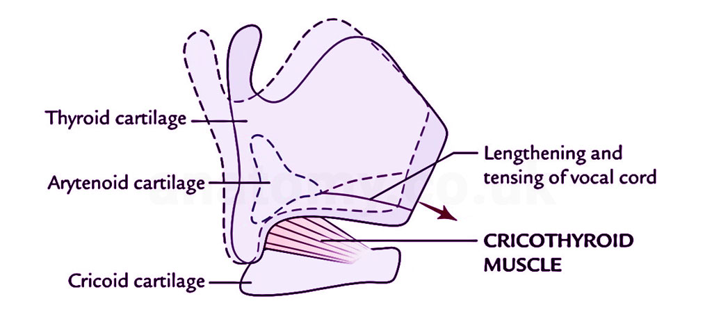
Image 2: Diagram of Cricothyroid joint
The cricothyroid joint, though modest in size, consists of several integral components that enable its complex function within the laryngeal anatomy.Articular Surfaces
The joint is formed by the articulation of two main surfaces:- The lateral aspect of the cricoid cartilage.
- The inferior cornu and the lower portion of the lamina of the thyroid cartilage.
Joint Capsule
The joint is enveloped by a fibrous capsule that is relatively thin but strong. This capsule is lined with synovial membrane, which secretes synovial fluid to reduce friction and ensure smooth movement within the joint.Ligamentous Support
Several ligaments contribute to the stability of the cricothyroid joint:- Anterior Cricothyroid Ligament: This ligament extends between the front parts of the cricoid and thyroid cartilages.
- Posterior Cricothyroid Ligament: This is situated at the back of the joint and is often thinner and less distinct than its anterior counterpart.
- Lateral Cricothyroid Ligaments: These ligaments, though less prominent, provide lateral stability to the joint.
Synovial Membrane
The inner lining of the joint capsule, the synovial membrane, produces synovial fluid. This fluid lubricates the joint, reducing friction between the articular surfaces during movement.Adjacent Musculature
The cricothyroid muscles, located laterally to the joint, play a pivotal role in its function. By contracting, these muscles influence the movement of the thyroid cartilage relative to the cricoid cartilage, modulating vocal cord tension.Biomechanics of the Joint
 The cricothyroid joint, while being a small and localized articulation, exhibits a range of motions that are central to voice modulation. To understand its function, it's essential to delve into the biomechanical properties that underpin its movements.
Types of Movement
Pivotal Motion: This movement allows the thyroid cartilage to tilt anteriorly and inferiorly relative to the cricoid. As this happens, the distance between the thyroid cartilage and arytenoids increases, leading to the stretching and tensioning of the vocal cords. This pivotal action plays a vital role in raising the pitch of the voice. Gliding Motion: A limited side-to-side gliding motion can occur at the cricothyroid joint. While not as pronounced as the pivotal motion, this gliding helps in fine-tuning laryngeal adjustments.Muscular Influence
The cricothyroid muscle, attaching laterally to both the cricoid and thyroid cartilages, is the primary muscle affecting the biomechanics of the joint. When this muscle contracts, it exerts a force that pivots the thyroid cartilage anteriorly and inferiorly, facilitating the tensioning of the vocal cords.Stability and Restraint
The capsule and ligaments surrounding the cricothyroid joint, particularly the anterior and posterior cricothyroid ligaments, provide stability. They ensure that movements remain within physiological limits, preventing hyperextension or excessive rotation which could damage the vocal apparatus.Role in Voice Modulation
The biomechanics of the cricothyroid joint directly impact the tension of the vocal cords. As the joint allows for the anterior tilting of the thyroid cartilage, the vocal cords are stretched, increasing their tension and resulting in a higher pitch. Conversely, when the joint's movement is relaxed, the pitch is lowered.Role in Phonation
Phonation, the process of voice production, involves a complex interplay of various laryngeal structures. The cricothyroid joint plays a pivotal role in this intricate mechanism, particularly in modulating the pitch and tone of the voice.Tension Modulation
The primary function of the cricothyroid joint is to adjust the tension of the vocal cords. As the joint facilitates the tilting of the thyroid cartilage, the vocal cords, which are attached to the thyroid and arytenoid cartilages, are stretched. An increase in tension results in the vocal cords vibrating at a higher frequency, producing a higher pitch.Vocal Range
The flexibility and range of movement within the cricothyroid joint, in conjunction with the intrinsic muscles of the larynx, determine an individual's vocal range. The ability of this joint to pivot and glide allows for a wide variation in vocal cord tension, contributing to the ability to produce a range of pitches.Fine-tuning of Voice
While the primary muscles responsible for tensioning the vocal cords are the cricothyroid muscles, the nuanced movements at the cricothyroid joint allow for delicate adjustments. This is especially important for activities that require precision in voice modulation, such as singing.Voice Quality and Resonance
The tension and length of the vocal cords, influenced by the cricothyroid joint's biomechanics, not only determine pitch but also play a role in voice quality and resonance. By altering cord tension, the joint indirectly influences the timbre and tonal qualities of the voice.Pathological Considerations
Any dysfunction or pathology affecting the cricothyroid joint can have ramifications on phonation. Conditions like arthritis or traumatic injuries that restrict the joint's movement can result in a limited vocal range or voice disorders.Pathologies and Clinical Considerations
The cricothyroid joint, while robust in its function, can be affected by various pathologies and disorders. Understanding these conditions and their clinical implications is crucial for effective diagnosis and intervention.- Arthritis: Like other joints in the body, the cricothyroid joint can be susceptible to arthritis, particularly rheumatoid arthritis. This inflammatory condition can result in pain, reduced mobility of the joint, and subsequently, voice changes or dysphonia.
- Traumatic Injuries: Direct trauma or forceful impact to the anterior neck can result in injury to the cricothyroid joint. This may manifest as joint dislocation, fracture of the adjacent cartilages, or damage to the supporting ligaments. Such injuries can be acute, causing immediate voice changes, pain, and swelling.
- Surgical Implications: In procedures involving the anterior larynx, such as tracheostomy or laryngeal surgeries, understanding the anatomy and biomechanics of the cricothyroid joint is paramount. Unintended damage to the joint can have lasting implications on voice and airway function.
- Tumors and Growths: Though less common, tumors or cysts can develop in proximity to the cricothyroid joint. Depending on their size and location, they might impede joint function or exert pressure on the vocal cords, leading to voice disturbances.
- Diagnostic Modalities: For assessing pathologies of the cricothyroid joint, various diagnostic tools can be employed. Laryngoscopy offers a direct visualization of the laryngeal structures, while imaging techniques like CT scans or MRIs provide detailed insights into joint anatomy and potential abnormalities.
- Therapeutic Interventions: Treatment for cricothyroid joint pathologies varies based on the underlying condition. Management can range from conservative approaches like voice therapy or physiotherapy to more invasive procedures like surgical interventions or injections.
Evolutionary and Comparative Anatomy
Delving into the evolutionary trajectory and comparative anatomy of the cricothyroid joint offers insights into its development over time and its role across various species.Evolutionary Significance
The evolution of complex vocalization in humans, and in many animals, is closely tied to the refinement of laryngeal structures. The cricothyroid joint, with its ability to modulate vocal cord tension, is believed to have played a role in the development of diverse vocal capabilities. [1]As early hominids evolved, the need for more nuanced communication could have driven the sophistication of this joint, supporting a wider range of vocal pitches.Comparison Across Mammals
In many mammals, the basic structure of the larynx, including components like the cricothyroid joint, is conserved. However, the degree of movement and function can vary. For instance:- In canines and felines, the joint allows for pitch modulation, though not as diverse as in humans.
- In some primates, the cricothyroid joint's function and range are more similar to humans, reflecting the need for varied vocal communications.
Birds and Vocalization
Birds, renowned for their vocal capabilities, possess a syrinx instead of a larynx. However, the principles of tension modulation for pitch control remain consistent, though the anatomical structures and their biomechanics differ.[2]Aquatic Mammals
Species like whales and dolphins, with their intricate vocalizations, have a laryngeal anatomy distinct from terrestrial mammals. While the basic components are recognizable, the function and mobility of joints like the cricothyroid may differ, catering to their unique vocal and respiratory needs in aquatic environments.Evolutionary Adaptations
Certain species have evolved unique adaptations related to the cricothyroid joint and its associated structures.[3] These adaptations, whether for specific vocal needs or environmental challenges, highlight the joint's significance in communication and survival. In examining the cricothyroid joint from an evolutionary and comparative lens, we appreciate its role not just in human phonation but also in the broader tapestry of animal communication and adaptation.Frequently Asked Questions
What is the primary function of the cricothyroid joint?
The cricothyroid joint's primary function is to adjust the tension of the vocal cords. By facilitating movements of the thyroid cartilage relative to the cricoid cartilage, it plays a crucial role in modulating the pitch of the voice.[4]How does the cricothyroid joint influence voice pitch?
The joint allows the thyroid cartilage to tilt, which in turn stretches the vocal cords. When the vocal cords are stretched and tensioned, they produce a higher pitched sound during vibration. Conversely, relaxed movements result in a lower pitch.[5]Can issues with the cricothyroid joint affect voice quality?
Yes. Pathologies or dysfunctions of the cricothyroid joint can lead to voice disturbances. Conditions such as arthritis, traumatic injuries, or tumors can impact the joint's mobility, potentially resulting in voice changes or dysphonia.How is the cricothyroid joint different from other laryngeal joints?
The larynx has two primary synovial joints: the cricothyroid and the cricoarytenoid joints.[6] While the cricothyroid joint modulates vocal cord tension (and thereby pitch) by adjusting the position of the thyroid cartilage, the cricoarytenoid joint is primarily involved in opening and closing the glottis by rotating the arytenoid cartilages.Are there specific exercises or therapies for the cricothyroid joint?
Voice therapy often incorporates exercises that indirectly target the functionality of the cricothyroid joint.[7] These exercises aim to improve voice quality, pitch control, and overall vocal health. However, direct interventions or therapies for the joint itself are usually considered in the context of specific pathologies or dysfunctions.Published on October 9, 2023
Last updated on April 24, 2025
Last updated on April 24, 2025