E
Epiglottis
Flap of cartilage that covers trachea while swallowing
RegionHead and Neck
SystemRespiratory System
What is Epiglottis?
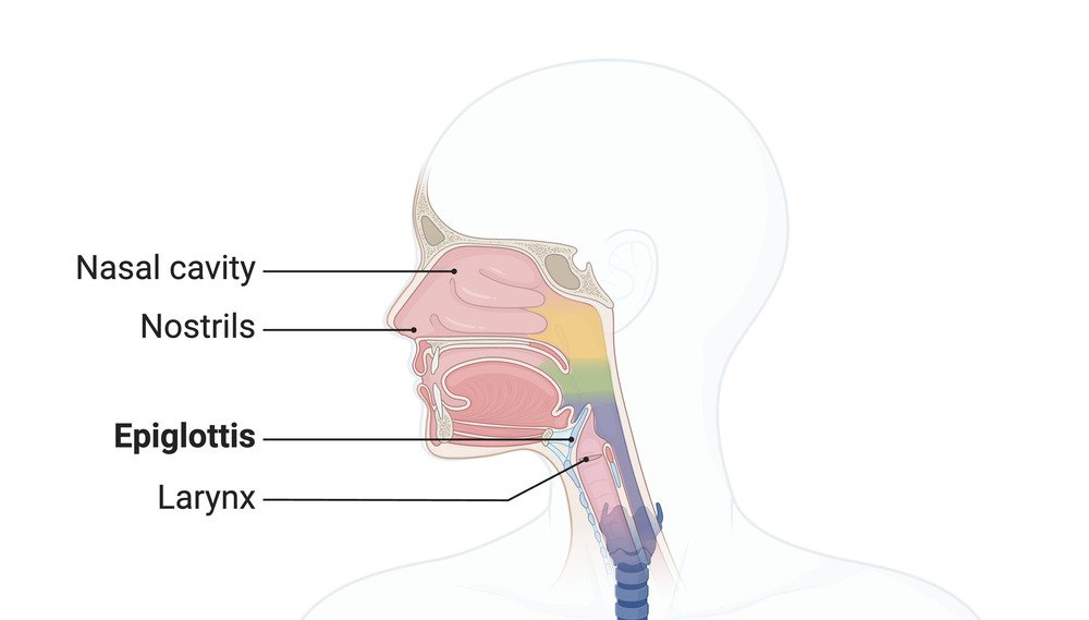
The epiglottis is a thin, leaf-shaped flap of cartilage located at the base of the tongue. It is covered with a mucous membrane and plays a critical role in separating the respiratory and digestive tracts. Made primarily of elastic cartilage, the epiglottis is flexible and can move to cover the opening of the larynx during swallowing.[2] This prevents food and liquids from entering the airway, ensuring they are directed into the esophagus instead. Its structure allows it to act as a protective barrier for the respiratory system.
Where is it Located?
The epiglottis is located in the throat, just behind the root of the tongue and above the larynx (voice box).[3] It is attached to the thyroid cartilage and connected to the hyoid bone, positioned at the entrance of the larynx.Anatomy
Shape and Structure
The epiglottis is a leaf-shaped structure composed primarily of elastic cartilage, making it both flexible and resilient. It has two distinct parts:- Superior (Lingual) Surface – The upper, free surface faces the tongue and oropharynx.
- Inferior (Laryngeal) Surface – The lower surface is directed towards the laryngeal inlet.
Cartilage Composition
The main structural component of the epiglottis is elastic cartilage, which provides flexibility and maintains its shape even after bending. Unlike hyaline cartilage, elastic cartilage contains elastin fibers, which enable repeated movements without deformation.Attachments
- To the Thyroid Cartilage – The epiglottis is anchored at its base (petiolus) to the posterior aspect of the thyroid cartilage through the thyroepiglottic ligament.
- To the Hyoid Bone – It is connected to the hyoid bone via the hyoepiglottic ligament, which maintains its position relative to the hyoid.
- To the Tongue – Three mucosal folds, called glossoepiglottic folds (one median and two lateral), attach the epiglottis to the posterior surface of the tongue.
Epithelium Covering
The epiglottis is covered by two types of epithelial tissue:- Non-keratinized Stratified Squamous Epithelium – Found on the upper (lingual) surface, providing protection against mechanical and chemical irritation during swallowing.
- Pseudostratified Ciliated Columnar Epithelium – Found on the lower (laryngeal) surface, which aids in mucus production and clearing debris.[7]
Mucosa and Glands
The mucosa of the epiglottis contains mucous glands, especially concentrated in the posterior surface. These glands secrete mucus to keep the epiglottis moist and lubricated, reducing friction during swallowing.Ligaments and Folds
- Glossoepiglottic Folds – Connect the epiglottis to the tongue.
- Aryepiglottic Folds – Extend from the epiglottis to the arytenoid cartilages, forming part of the laryngeal inlet.
- Hyoepiglottic Ligament – Attaches the anterior surface of the epiglottis to the hyoid bone.
- Thyroepiglottic Ligament – Secures the epiglottis to the thyroid cartilage.
Vascular Supply
The blood supply to the epiglottis comes primarily from branches of the superior laryngeal artery, a branch of the superior thyroid artery, which itself arises from the external carotid artery. Venous drainage mirrors the arterial supply, draining into the internal jugular vein.[8]Innervation
The epiglottis receives sensory innervation from the internal branch of the superior laryngeal nerve, a branch of the vagus nerve (Cranial Nerve X). This nerve supplies both sensation and reflex pathways essential for its protective actions.Lymphatic Drainage
Lymphatic drainage of the epiglottis primarily occurs via the deep cervical lymph nodes, which are involved in clearing lymph from the surrounding tissues.[1]Microscopic Anatomy
- Elastic Cartilage Framework – Provides flexibility and resilience.
- Glandular Tissues – Mucous glands embedded within connective tissue.
- Lamina Propria – A layer of loose connective tissue beneath the epithelium, rich in blood vessels and immune cells.
Function
Airway Protection During Swallowing
The primary function of the epiglottis is to act as a protective barrier for the airway during swallowing (deglutition).- Mechanism of Action:
- Coordinated Movements:
- The elevation of the larynx is facilitated by the hyoid bone and associated muscles.
- The tongue pushes the bolus (food mass) toward the pharynx, initiating the reflexive closure of the epiglottis.[4]
Separation of Respiratory and Digestive Pathways
The epiglottis functions as a gatekeeper between the respiratory tract and the digestive tract:- It ensures that air passes through the larynx into the trachea during breathing.
- It directs food and liquids into the esophagus during swallowing.
Cough Reflex Activation
The epiglottis plays a key role in triggering the cough reflex:- If any foreign particle or liquid mistakenly enters the laryngeal inlet, sensory receptors in the epiglottis detect the intrusion.
- This signals the vagus nerve (Cranial Nerve X), prompting a cough reflex to expel the substance and protect the airway.
Voice Modulation
The epiglottis indirectly assists in voice modulation:- It contributes to the resonance and tone of the voice by influencing airflow through the larynx.
- During speech, the movement of the epiglottis alters the shape of the vocal tract, affecting sound production.
Maintaining Airway Patency During Breathing
When not swallowing, the epiglottis remains in an upright, open position to allow:- Uninterrupted airflow from the pharynx to the trachea and lungs.
- Optimal oxygen exchange without obstruction.
Moistening and Lubricating the Throat
- The mucous glands on the surface of the epiglottis produce mucus to keep it moist and lubricated.
- This reduces friction during swallowing and allows smooth movement of food and liquids.
Reflexive Movements
The epiglottis performs reflexive movements controlled by the autonomic nervous system, ensuring involuntary actions such as:- Closing over the glottis during swallowing.
- Returning to its resting position after the act of swallowing.[6]
Preventing Aspiration During Vomiting
The epiglottis also plays a role in protecting the airway during episodes of vomiting:- It closes to ensure stomach contents do not enter the trachea.
- This helps prevent choking and aspiration of vomit into the lungs.
Sensory and Immune Response
- Sensory Role: The epiglottis has sensory nerve endings that can detect irritation and trigger protective reflexes.
- Immune Defense: It contributes to the mucosal immune system by trapping pathogens and particles in its mucus layer, preventing their entry into the respiratory tract.
Clinical Significance
The epiglottis plays a critical role in protecting the airway, and any dysfunction can lead to serious health issues. Epiglottitis is one of the most notable conditions, characterized by inflammation and swelling of the epiglottis, often caused by bacterial infections like Haemophilus influenzae type b (Hib). This condition can lead to airway obstruction, making it a medical emergency requiring immediate attention. Aspiration is another concern, where food or liquids enter the airway due to improper closure of the epiglottis, potentially causing aspiration pneumonia. Trauma, burns, or intubation injuries can also damage the epiglottis, affecting its function. Structural abnormalities, such as laryngomalacia, may result in floppy epiglottis tissue that obstructs airflow, especially in infants. Evaluation of epiglottis-related issues often involves imaging studies like laryngoscopy or CT scans, and treatments range from antibiotics and steroids to emergency airway management, including tracheostomy in severe cases.Published on January 7, 2025
Last updated on May 11, 2025
Last updated on May 11, 2025